Adaptive optics meets AI for cellular-scale eye care
Retinal biomarkers reveal systemic risks, linking eye imaging to cardiometabolic disease management.
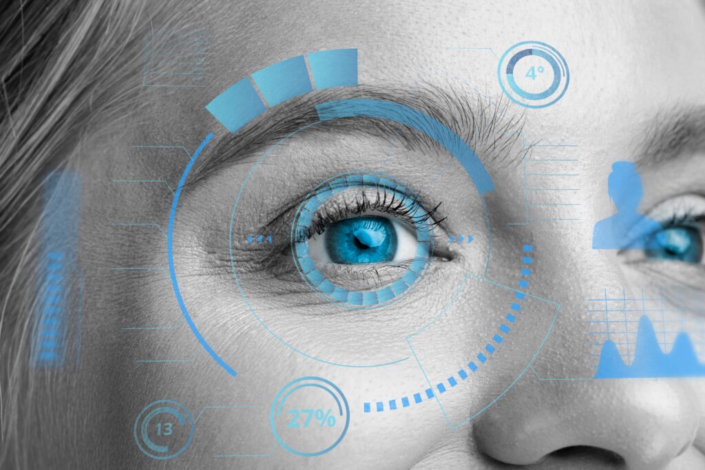
AI is moving from lab demos to frontline eye care, with clinicians using algorithms alongside routine fundus photos to spot disease before symptoms appear. The aim is simple: catch diabetic retinopathy early enough to prevent avoidable vision loss and speed referrals for treatment.
New imaging workflows pair adaptive optics with machine learning to shrink scan times from hours to minutes while preserving single-cell detail. At the US National Eye Institute, models recover retinal pigment epithelium features and clean noisy OCT data to make standard scans more informative.
Duke University’s open-source DCAOSLO goes further by combining multiplexed light signals with AI to capture cellular-scale images quickly. The approach eases patient strain and raises the odds of getting diagnostic-quality data in busy clinics.
Clinic-ready diagnostics are already changing triage. LumineticsCore, the first FDA-cleared AI to detect more-than-mild diabetic retinopathy from primary-care images, flags who needs urgent referral in seconds, enabling earlier laser or pharmacologic therapy.
Researchers also see the retina as a window on wider health, linking vascular and choroidal biomarkers to diabetes, hypertension and cardiovascular risk. Standardised AI tools promise more reproducible reads, support for trials and, ultimately, home-based monitoring that extends specialist insight beyond the clinic.
Would you like to learn more about AI, tech, and digital diplomacy? If so, ask our Diplo chatbot!