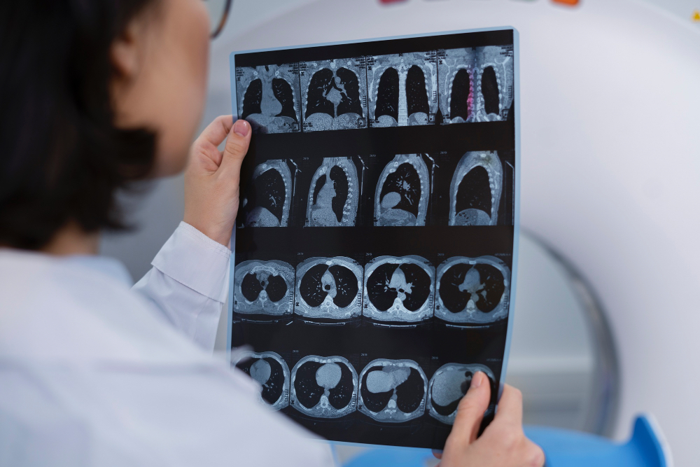
AI brain atlas reveals unprecedented detail in MRI scans
Created from high-resolution post-mortem data, NextBrain combines AI and MRI for unprecedented anatomical detail in living patients.

Researchers at University College London have developed NextBrain, an AI-assisted brain atlas that visualises the human brain in unprecedented detail. The tool links microscopic tissue imaging with MRI, enabling rapid and precise analysis of living brain scans.
NextBrain maps 333 brain regions using high-resolution post-mortem tissue data, which is combined into a digital 3D model with the aid of AI. The atlas was created over the course of six years by dissecting, photographing, and digitally reconstructing five human brains.
AI played a crucial role in aligning microscope images with MRI scans, ensuring accuracy while significantly reducing the time required for manual labelling. The atlas detects subtle changes in brain sub-regions, such as the hippocampus, crucial for studying diseases like Alzheimer’s.
Testing on thousands of MRI scans demonstrated that NextBrain reliably identifies brain regions across different scanners and imaging conditions, enabling detailed analysis of ageing patterns and early signs of neurodegeneration.
All data, tools, and annotations are openly available through the FreeSurfer neuroimaging platform. The public release of NextBrain aims to accelerate research, support diagnosis, and improve treatment for neurological conditions worldwide.
Would you like to learn more about AI, tech and digital diplomacy? If so, ask our Diplo chatbot!
