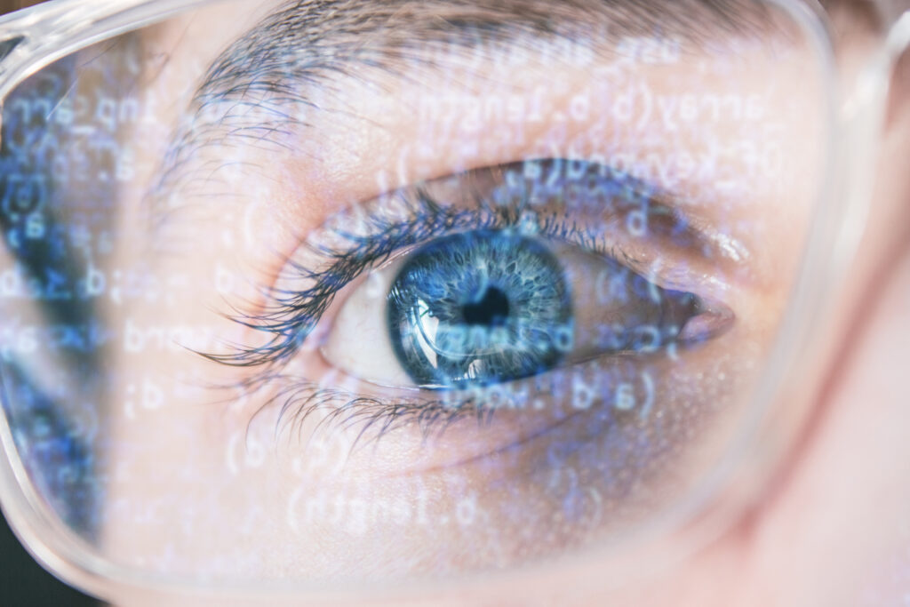
AI transforms retinal scans into predictive health tools
Deep learning frameworks can now estimate blood sugar levels directly from retinal images, reducing the need for traditional tests.

AI and machine learning are transforming ophthalmology, making retinal scans powerful tools for predicting health risks. Retinal images offer a non-invasive view of blood vessels and nerve fibres, showing risks for high blood pressure, kidney, heart, and stroke-related issues.
Technologies like fundus photography and optical coherence tomography-angiography (OCT-A) now enable detailed imaging of retinal vessels. Researchers use AI to analyse these images, identifying microvascular biomarkers linked to systemic diseases.
Novel approaches such as ‘oculomics’ allow clinicians to predict surgical outcomes for macular hole treatment and assess patients’ risk levels for multiple conditions in one scan.
AI is also applied to diabetes screening, particularly in countries with significant at-risk populations. Deep learning frameworks can estimate average blood sugar levels (HbA1c) from retinal images, offering a non-invasive, cost-effective alternative to blood tests.
Despite its promise, AI in ophthalmology faces challenges. Limited and non-diverse datasets can reduce accuracy, and the ‘black box’ nature of AI decision-making can make doctors hesitant.
Collaborative efforts to share anonymised patient data and develop more transparent AI models are helping to overcome these hurdles, paving the way for safer and more reliable applications.
Would you like to learn more about AI, tech and digital diplomacy? If so, ask our Diplo chatbot!
